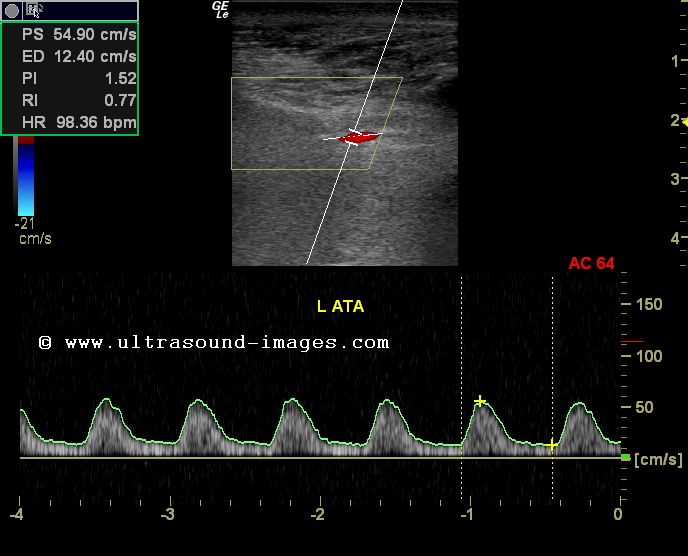
Within two days a copy of the test results will be sent to the ordering provider. Compression ultrasonography with or without Doppler waveform analysis has a high sensitivity (95) and specificity (96) for proximal thrombosis however, the sensitivity is lower for calf veins.


The physician will interpret the images and velocity measurements. The technologist is not permitted to discuss the results with you. You will be able to leave as soon as the test is completed. Ultrasonography combined with Doppler imaging has been found to useful in the mapping of the vascular anatomy of lower limbs. These exams will take 30-45 minutes to complete. PATIENT PREPARATION: Introduce yourself to patient. The technologist performing the exam is specially trained to evaluate the lower extremity arteries for abnormalities, and will create Doppler waveforms and pressure measurements of the vessels in the legs. Duplex ultrasound with color flow Doppler with transducer frequencies ranging from 3.5-10. This is to measure the amount of blood flow post-exercise and the recovery time needed. In this particular study the patient walks on a treadmill for 5 minutes and then is asked to lie back on the stretcher where blood pressure cuffs are placed on the ankle. The rationale for exercising patients in the vascular laboratory is to replicate the symptoms the patient experiences outside of the lab and to document the effects. Many patients with PAD, however, experience symptoms after some sort of exercise. Non-invasive studies done on a patient in a resting state provide information about the vascular system in a “non-stressed” condition. Peripheral Arterial disease (PAD) refers to narrowing of the blood vessels, which obstructs blood flow to the legs, usually due to atherosclerosis (fatty plaque in walls of blood vessels) or other diseases. Your healthcare provider has ordered a Lower Extremity Arterial examination.


 0 kommentar(er)
